Discovering a new bump anywhere on your body can be alarming, especially if it appears suddenly and has a distinct appearance. A raised, waxy-looking bump on your temple might not only be concerning due to its unexpected presence but also because of its location on your face, where it is readily visible. While it could be benign, understanding what it might be and when to seek medical advice is crucial.
In these uncertain times, getting an immediate appointment with a dermatologist might not always be possible. However, arming yourself with knowledge can ease anxieties and help you make informed decisions about your skin health. This article aims to shed light on the nature of these bumps, focusing on potential causes such as seborrheic keratosis, and guiding you on safe interim care while you await professional evaluation.
1. Why a New Waxy Bump on Your Temple Deserves a Closer Look
Any new skin growth warrants attention, particularly if it arises on your face. A waxy, raised bump could be a seborrheic keratosis, a common benign skin growth, but it could also mimic more serious conditions such as basal cell carcinoma or melanoma, which require prompt medical intervention.
These bumps can vary in size, often starting small, around 1-2 millimeters, and can grow larger over time, sometimes reaching several centimeters. Monitoring any changes in size, color, or texture is essential, as rapid or unusual changes might indicate a need for urgent medical assessment.
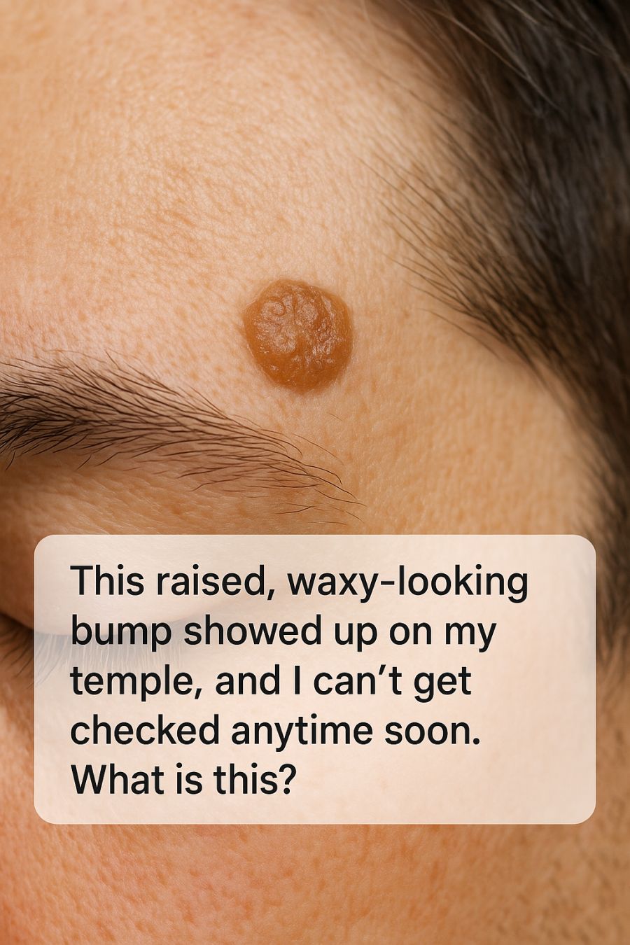
2. What Seborrheic Keratosis Is and Why It Looks So ‘Stuck On’
Seborrheic keratosis is a non-cancerous skin growth that typically appears in adulthood. Its hallmark feature is its ‘stuck-on’ appearance, often resembling a drop of wax on the skin. These growths are composed of keratinocytes, which are the predominant cells in the epidermis.
The waxy appearance is due to the accumulation of keratin on the skin’s surface. While they can appear anywhere on the body, they are most common on the face, chest, shoulders, and back. Seborrheic keratoses can range in color from light tan to dark brown or black, and they are usually elevated above the skin surface.
3. Classic Signs: Color, Texture, and Shape of Seborrheic Keratoses
Seborrheic keratoses are known for their distinctive appearance: they are often round or oval and have a well-defined border. Their texture can be smooth or slightly rough, and they often appear scaly or wart-like.
Color can vary significantly, with some lesions appearing yellowish, while others are a deep brown or black. Despite this variation, the consistency of their waxy, ‘pasted-on’ look is a key identifier, distinguishing them from other skin conditions. Keeping an eye on these attributes can help differentiate them from more concerning lesions.
4. How Seborrheic Keratosis Differs From Skin Cancer
While seborrheic keratoses are benign, they can sometimes mimic the appearance of melanoma or other skin cancers. The primary difference lies in their growth pattern and texture. Skin cancers often change in size, color, and shape more rapidly than benign growths.
Melanomas, for instance, might exhibit asymmetry, irregular borders, and multiple colors, including shades of red or blue. Seborrheic keratoses, conversely, maintain a consistent appearance over time, though any change in characteristics should still be evaluated by a healthcare professional to rule out malignancy.
5. When a ‘Benign’ Bump Still Needs a Doctor’s Opinion
Even though seborrheic keratosis is benign, consulting a doctor is crucial for a definitive diagnosis, especially if the bump is new, changing, or symptomatic. Symptoms such as itching, bleeding, or pain can indicate complications or misdiagnosis.
Dermatologists can often diagnose these growths with a visual examination but may perform a biopsy if there is any doubt about the nature of the lesion. This ensures that any potential malignancies are caught early, allowing for prompt and effective treatment.
6. Common Triggers: Age, Genetics, Sun Exposure, and Hormones
Seborrheic keratoses are most common in people over the age of 50, although younger adults can develop them as well. Genetics play a significant role, as these growths often run in families.
Sun exposure is another contributing factor, as it can accelerate the growth of seborrheic keratoses, especially in sun-exposed areas like the face and arms. Hormonal changes, too, can influence their development, particularly during pregnancy or periods of hormonal flux.
7. Why the Temple and Face Are Hotspots for These Growths
The face, including the temple area, is frequently exposed to sunlight, making it a common site for seborrheic keratoses. The thin skin in these regions may also be more susceptible to the factors that promote the growth of these lesions.
Additionally, the face has a high density of sebaceous glands and hair follicles, which may contribute to the proliferation of keratinocytes, the cells involved in forming seborrheic keratoses.
8. Viral Home Hacks and Why You Shouldn’t Pick, Peel, or Burn It Off
The internet is rife with home remedies for removing skin growths, ranging from apple cider vinegar to duct tape. However, such methods are not recommended for seborrheic keratosis, as they can lead to irritation, infection, or scarring.
Picking or peeling at these lesions can not only damage the skin but also complicate a future medical assessment. It is safer to wait for a professional evaluation and treatment plan, which minimizes the risk of adverse outcomes.
9. Safe At-Home Care While You Wait for an Appointment
While waiting for a medical appointment, there are steps you can take to care for your skin. Keeping the area clean and moisturized can help prevent irritation. Using a gentle cleanser and a fragrance-free moisturizer is advisable.
Avoiding sun exposure by wearing a wide-brimmed hat or using sunscreen can also prevent further irritation or growth. If the lesion is itchy, applying a cool compress or using a dermatologist-recommended anti-itch cream may provide relief.
10. Medical Treatments: Freezing, Shaving, Lasers, and Creams
Once you see a dermatologist, they might suggest several options for removing seborrheic keratoses. Cryotherapy, or freezing, is a common method that involves applying liquid nitrogen to the growth, causing it to fall off over time.
Shave excision is another option, where the lesion is carefully shaved off under local anesthesia. Laser therapy can also be effective, using targeted light beams to vaporize the growth. In some cases, topical treatments that contain alpha hydroxy acids or urea may help in softening and eventually removing the growth.
11. Red-Flag Symptoms That Mean You Shouldn’t Wait
Certain symptoms should prompt an immediate consultation with a dermatologist. These include rapid changes in size, irregular or blurred borders, multiple colors within the same lesion, or any accompanying symptoms such as bleeding, itching, or pain.
If the bump has a diameter larger than 6 millimeters (about the size of a pencil eraser) or if it is evolving in any way, it should be evaluated promptly to rule out skin cancer.
12. How to Document Changes: Photos, Skin Diaries, and Telehealth
Keeping a record of your skin changes can be very helpful for both you and your healthcare provider. Taking regular photos of the lesion with a date stamp can help track any changes over time.
Maintaining a skin diary to note any new symptoms or changes in existing lesions can also provide valuable information. If an in-person visit is not possible, telehealth services can offer a preliminary assessment and guidance based on your documentation.
13. Protecting Your Skin Going Forward: Sunscreen, Checks, and Routine Exams
Preventive care is crucial in maintaining healthy skin and minimizing the risk of seborrheic keratoses and other skin conditions. Regular use of a broad-spectrum sunscreen with an SPF of at least 30 can protect against harmful UV rays that contribute to skin changes.
Routine skin checks, either self-examinations or professional evaluations, are essential for early detection of any new or changing lesions. Scheduling regular dermatological exams can help ensure that any concerning growths are identified and treated promptly.
